CONTENT
- 01
Introduction
- 02
Requirements
- 03
Preparation
- 04
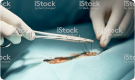
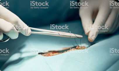
Procedure
- 05
Aftercare
- 06
Outcomes
- 07
Further Reading
Peridontal Flap Procedure
CANIS
INTRODUCTION
To insert a catheter into the cephalic vein as an intravenous route for the administration of drugs, fluids, etc or for blood sampling.
USES
- Fluid therapy Fluid therapy
- Anesthesia General anesthesia: overview
- Anesthetic induction Anesthetic induction: overview.
- Drug administration Therapeutics: antimicrobial drug Analgesia: overview
- Blood sampling Jugular venipuncture.
- Euthanasia.
ADVANTAGES
- Easily accessible, large size vein
- Easily performed in the conscious animal with simple restraint.
- Length of forearm allows simple secure fixing of catheter for semi-permanence.
- Bilateral sites available, ie both forelimbs.
DISADVANTAGES
- The cephalic vein can have a tortuous course in the brachicephalic breeds making location and fixation of the vein for catheterization difficult.
- The site is easily accessible for interference by the patient - catheter must be secured adequately and interference prevented, eg using an Elizabethan collar or muzzle.
DECISION TAKING
Criteria for choosing test
- The primary objective in treating periodontal disease Peridontal disease is to achieve a mouth free from pain and infection. Extraction of teeth ispreferable to leaving teeth in a diseased state.
- Is the disease localised or generalised? Generalised disease carries a poor prognosis for long-term success than localised disease.
- Are there any uncontrollable systemic factors that may negatively affect outcome? Is there furcational involvement? This makes treatment and homecare much more challenging.
- Consider tooth functionally. Preservation of a mandibular canine is a higher priority than a first pre-molor.
- Do you have adequate equipment and training to carry out the procedure effectively?
REQUIREMENTS
Materials required
Minimum equipment
- Periosteal elevators in several sizes.
- Ultrasonic scaler plus tips suitable for sub-gingival use.
- Hand scaler
- Gracey curettes, eg #11-12, 13-14.
- Scalpel handle.
- Periodontal probe.
- Sharp explorer probe
- Adson tissue forceps
- Needle holders.
- Tungsten carbide and round diamond burs
- Tissue retractor
Minimum consumables
- No. 15 scapel blade
- Suture material, ideally absorbable monofilament, eg pologlecaprone 25 Suture materials.
PREPARATION
Pre-medication
- Consider multi-modal and pre-emptive analgesic protocols as gold standard.
- Dietary preparation
- Fast patient for 12 hours prior to general anesthesia to prevent reflux esophagitis.
- Site preparation
- Ensure a cuffed endotracheal tube us used, and a retrievable throat pack placed.
- Flush the mouth with 0.12% chlorhexidine solution Chlorhexidine.
- Perform supragingival scaling and polishing Dental scaling.
- Regional anesthesia (nerve blocks Local anesthesia: intraoral ) are strongly recommended.
- Full mouth probing and charting is mandatory.
- Radiography Dental adiography: overview required for any treatment areas. Full mouth radiography can be justified
- Proper lighting and magnification if helpful.
FURTHER READING
Publications
Refereed papers
- Recent references from PubMed and VetMedResource.
- Beckman B(2003) Mandibular incisor apically repositioned flap in the dog. J Vet Dent 20(4), 245-240 PubMed
Other sources of information
- Newman M G, Takeu H H, Klollevold P R, Carranza F R (2012) Carranza's Clinical Periodontology.St Louis: Elsevier Saunders.
- Holmstrom S E, Frost Fitch P, Eisher E R (2004) Veterinary Dental techniques for the Small Animal Practitioner. Philadepiphia:Saunders.
- Wiggs R B, Lobprise H B (1997) Veterinary Dentistry Principles and Practice. Philadelphia: Lippincott-Raven.
SAME SERIES
Peridontal Flap Procedure - 01
Peridontal Flap Procedure - 01
Peridontal Flap Procedure - 01
Peridontal Flap Procedure - 01
Peridontal Flap Procedure - 01
Peridontal Flap Procedure - 01
RELATED CONTENT
CanisArticle
Paranasal Sinus: Bone Flap Technique -
Standing Surgery
CanisArticle
Paranasal Sinus: Bone Flap Technique -
Standing Surgery
CanisArticle
Paranasal Sinus: Bone Flap Technique -
Standing Surgery
CanisArticle
Paranasal Sinus: Bone Flap Technique -
Standing Surgery
CanisArticle
Paranasal Sinus: Bone Flap Technique -
Standing Surgery
CanisArticle
Paranasal Sinus: Bone Flap Technique -
Standing Surgery
CanisArticle
Paranasal Sinus: Bone Flap Technique -
Standing Surgery